


Katarina
This utterly baffling skull was unveiled in 2026. There are reasonable doubts about its authenticity.
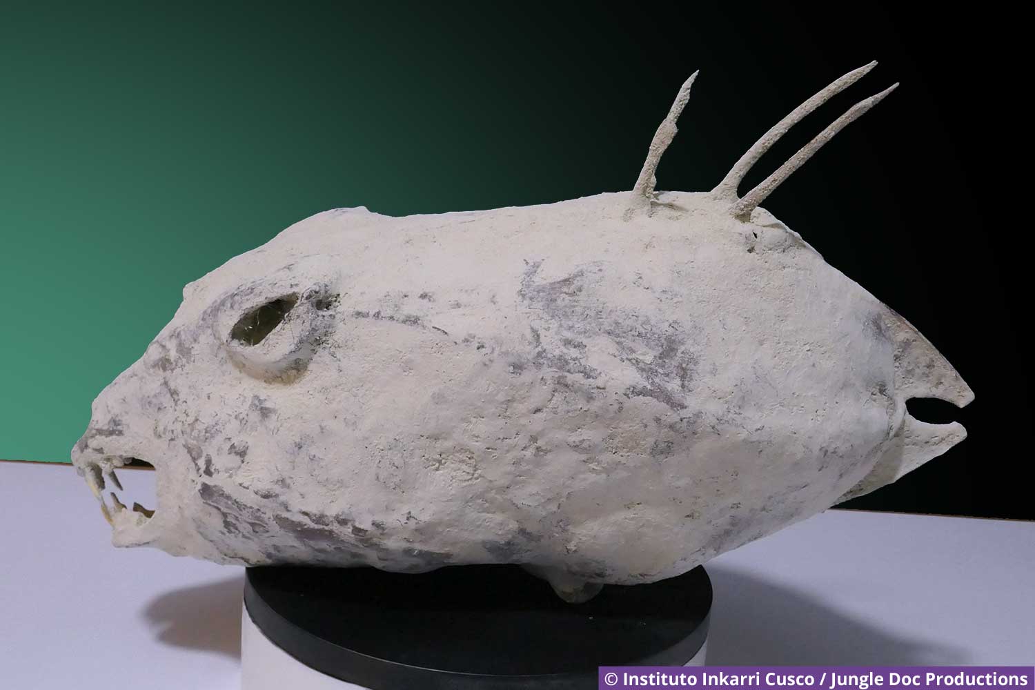
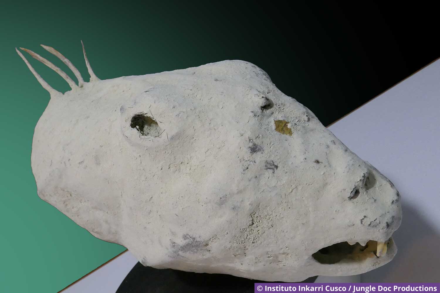
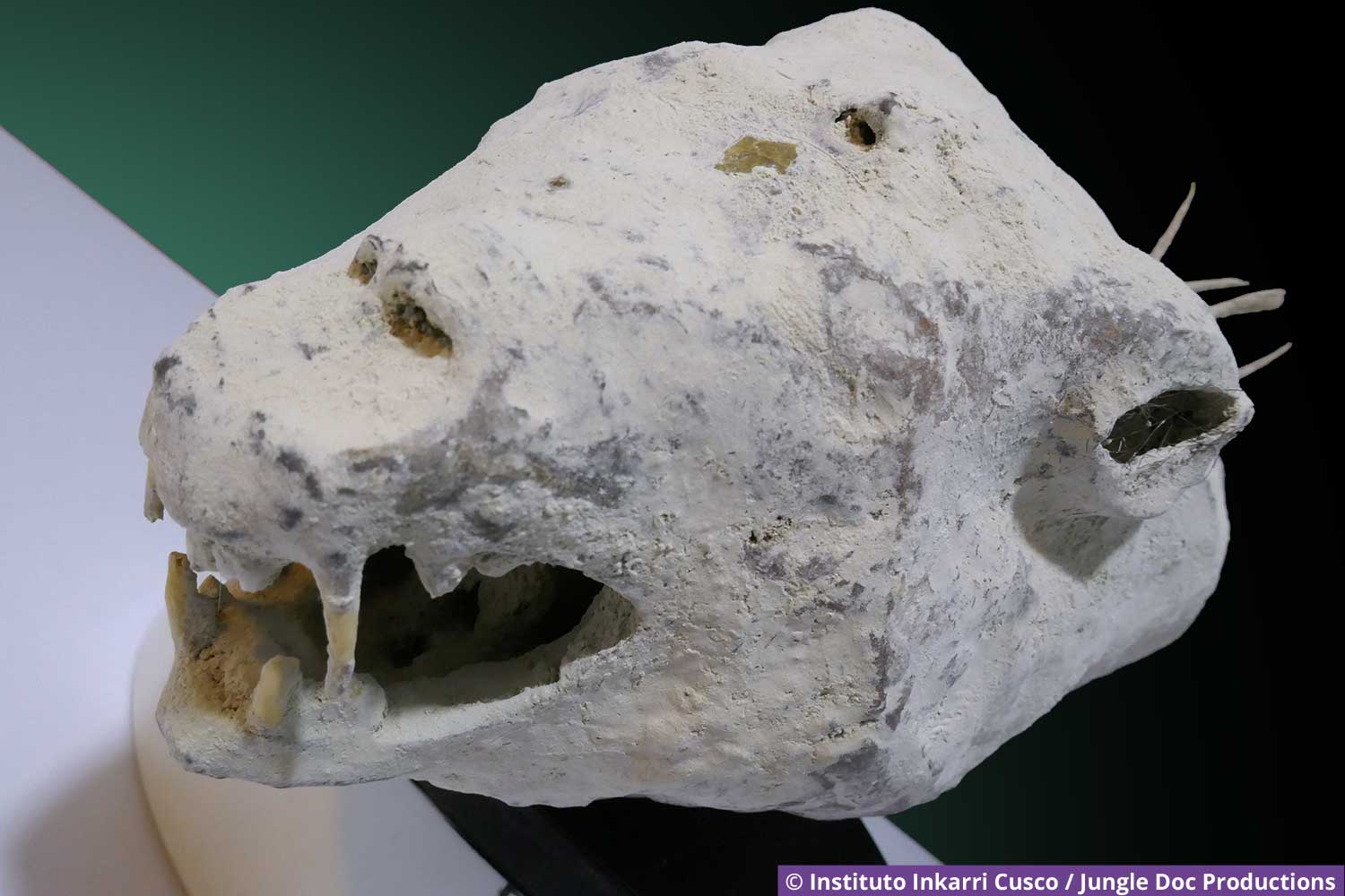
Katarina was discovered by Dr. Edson Salazar Vivenco.
Features
Length: 39 cm
Width: 15 cm
Height: 18 cm
Species: unknown
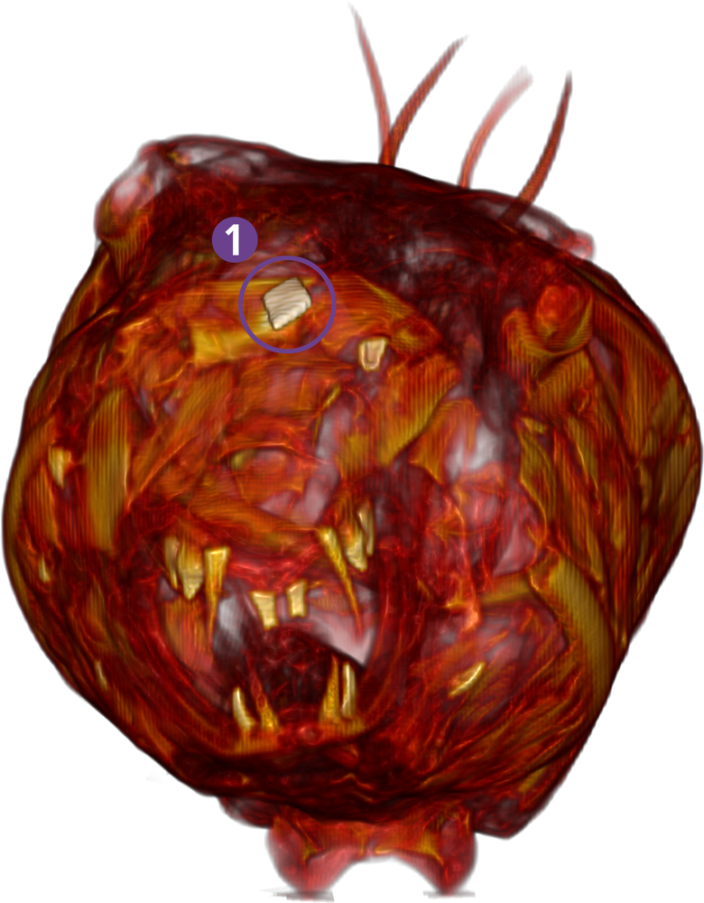
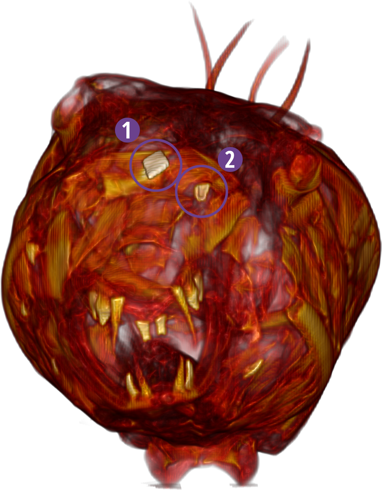
A metal implant on the head
and possibly an implant beneath the top layer
4 “spine”-shaped protrusions on the top of the rear section
Videos created by Frop-Frop based on the work of the DICOM
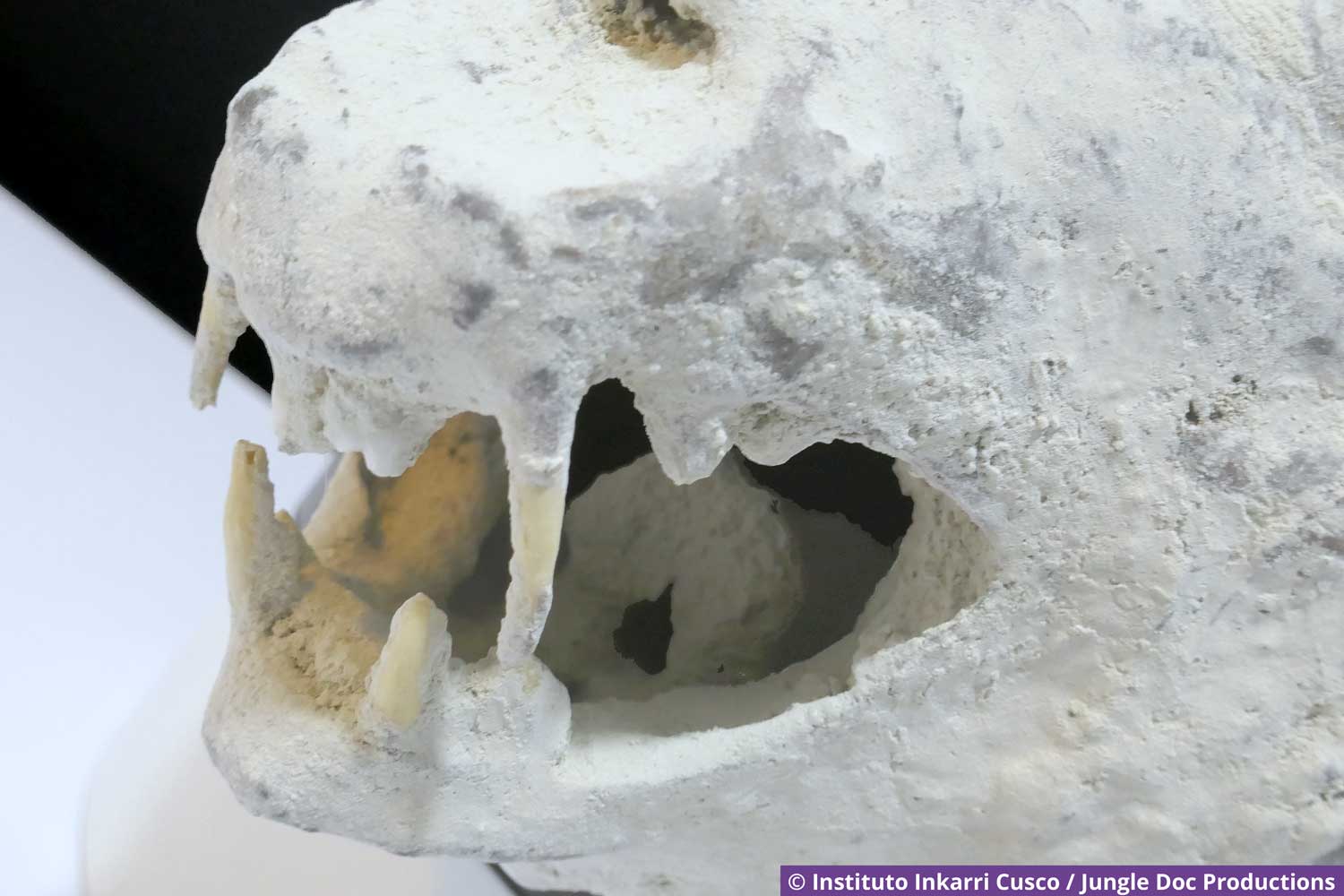
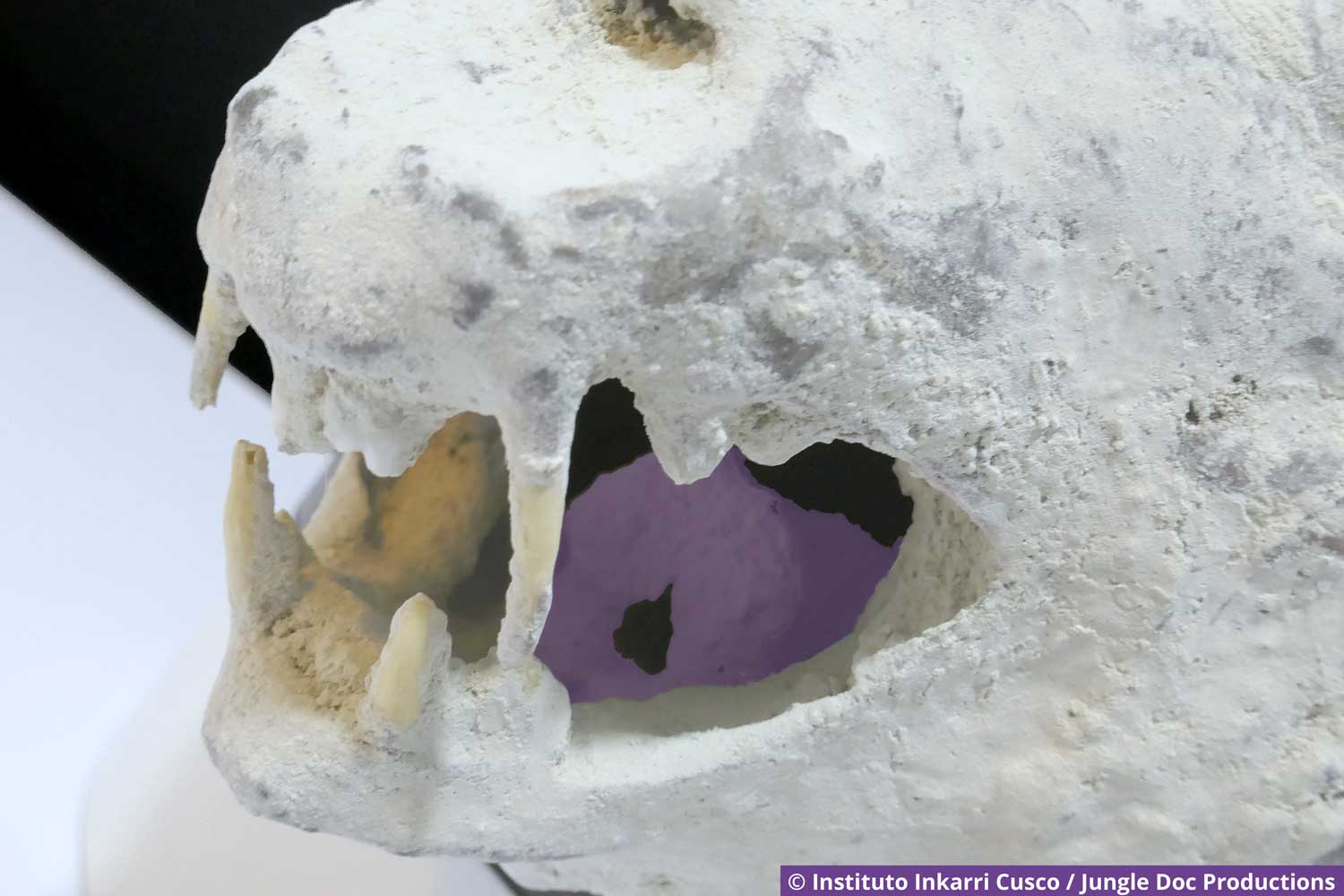
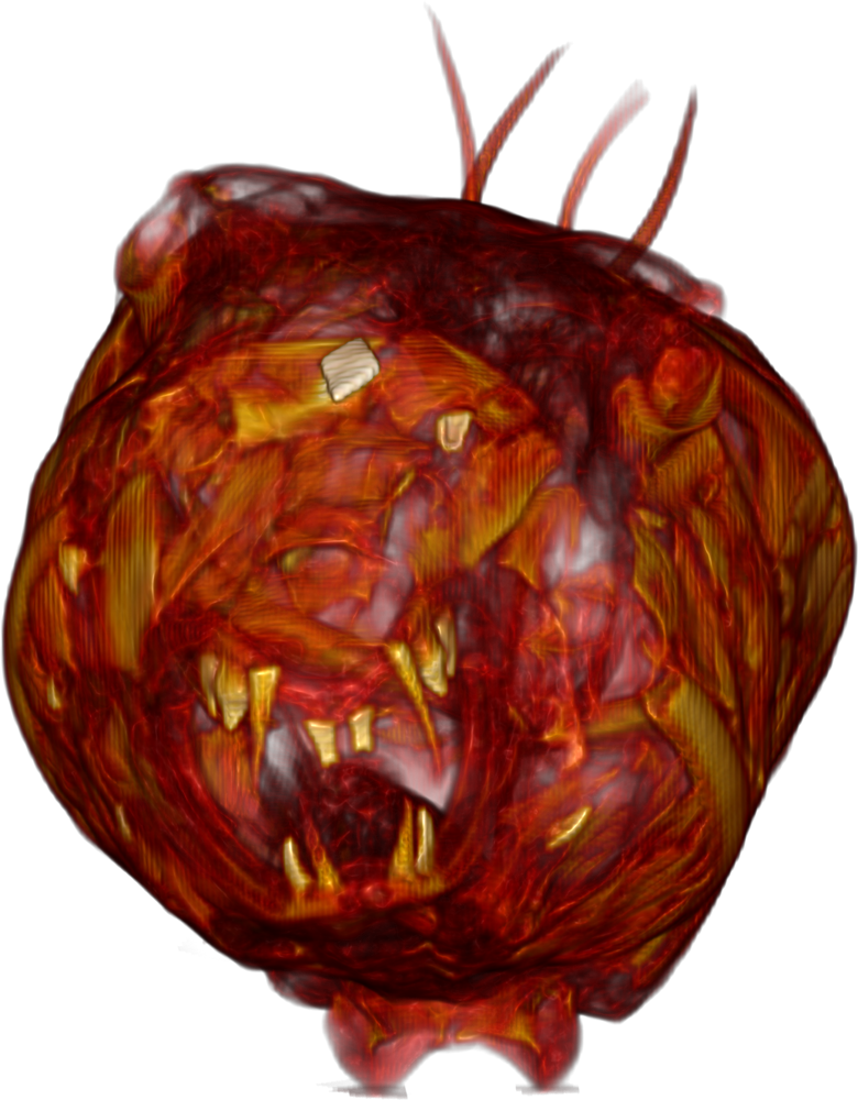
Katarina's dentition: 8 teeth on the upper jaw, 4 on the lower jaw. There is also what appears to be a *Jacobson's organ in the roof of her mouth.
*In snakes and lizards, this organ—also known as the “vomero-nasal organ”—is used to detect chemicals, for example, by collecting particles with their tongues and then depositing them in the organ.
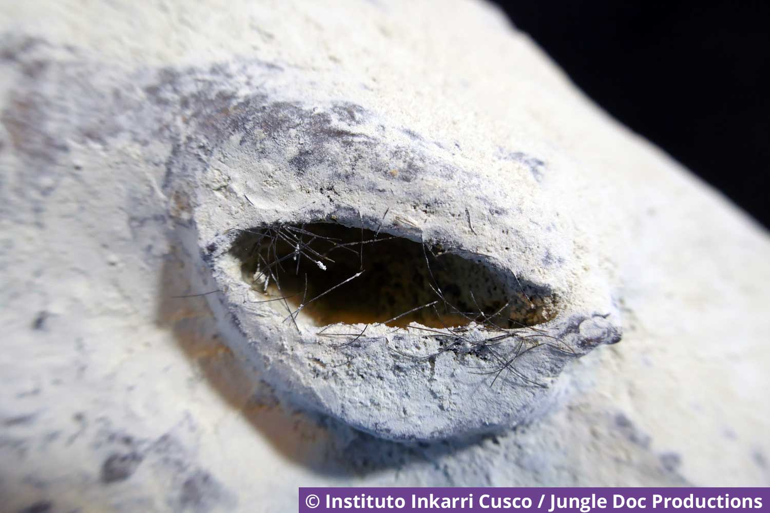
Katarina's right eye: you can see her eyelashes.
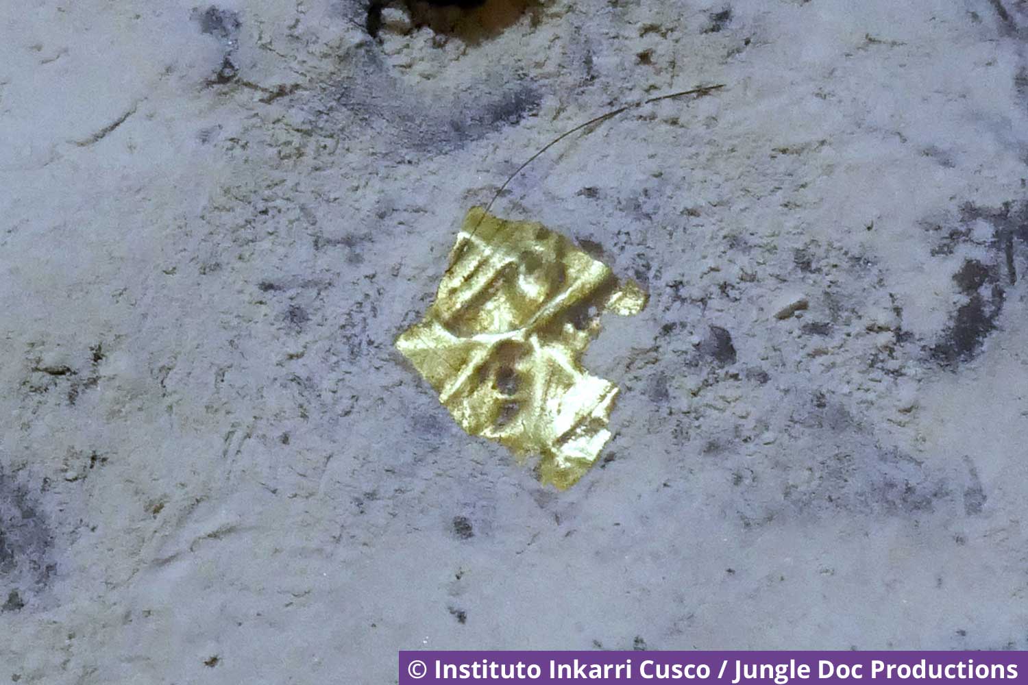
A gold-plated metal plate on Katarina's head
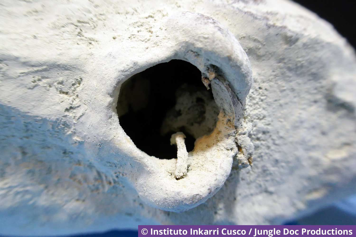
Katarina's foramen magnum
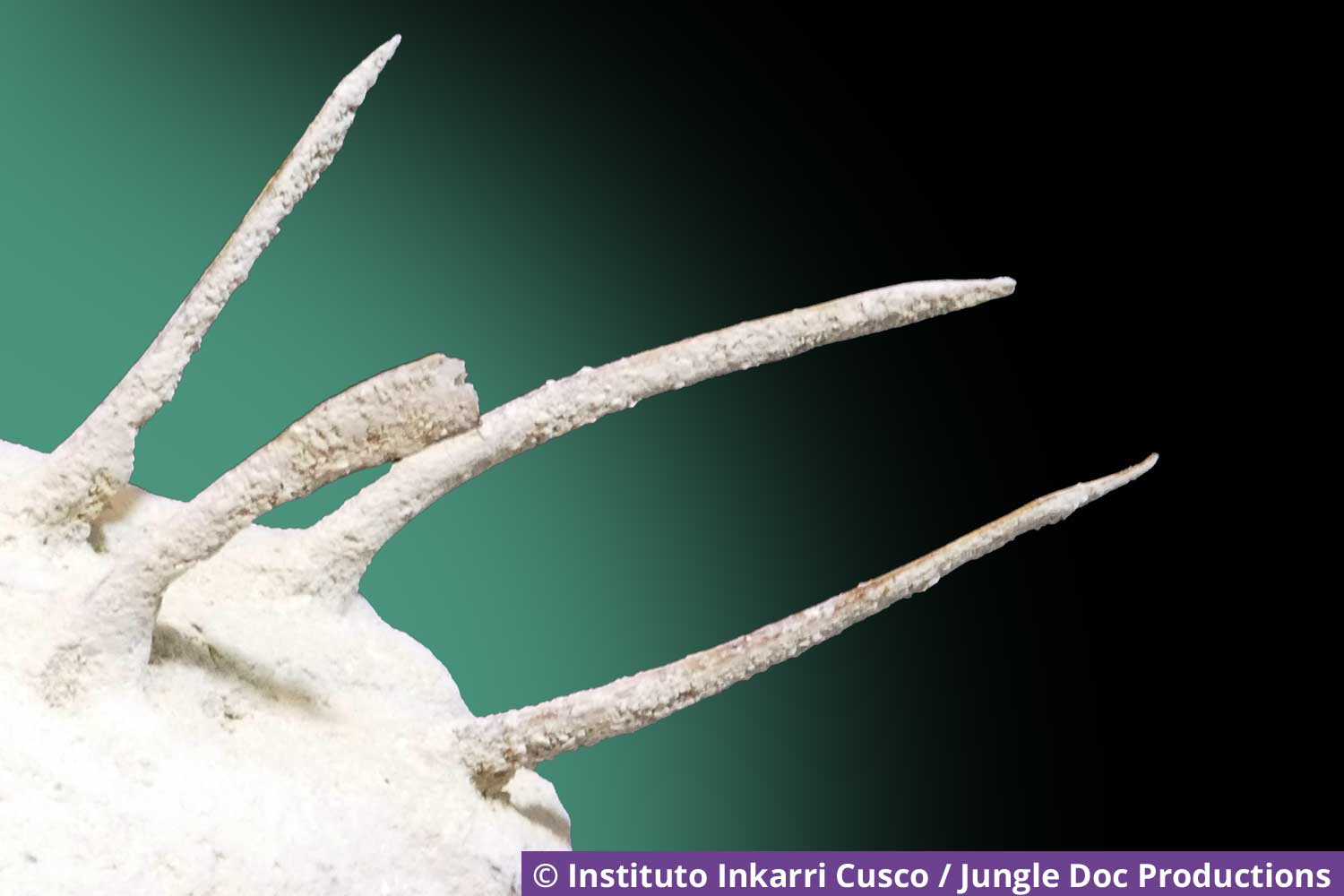
The “spines” on the top of the back of Katarina's skull. These are bony structures that contain soft tissue. They measure between 4 and 8 cm.
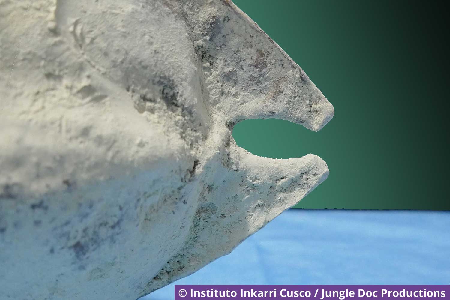
The back of Katarina's skull
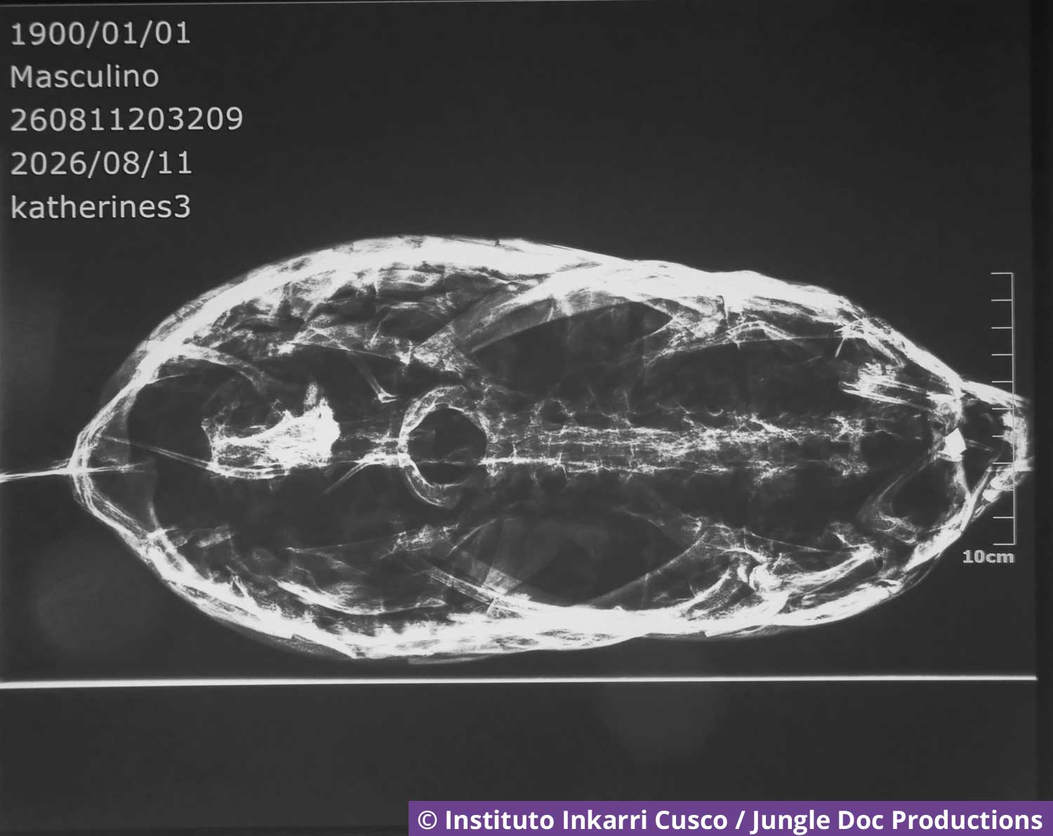
Katarina's X-rays
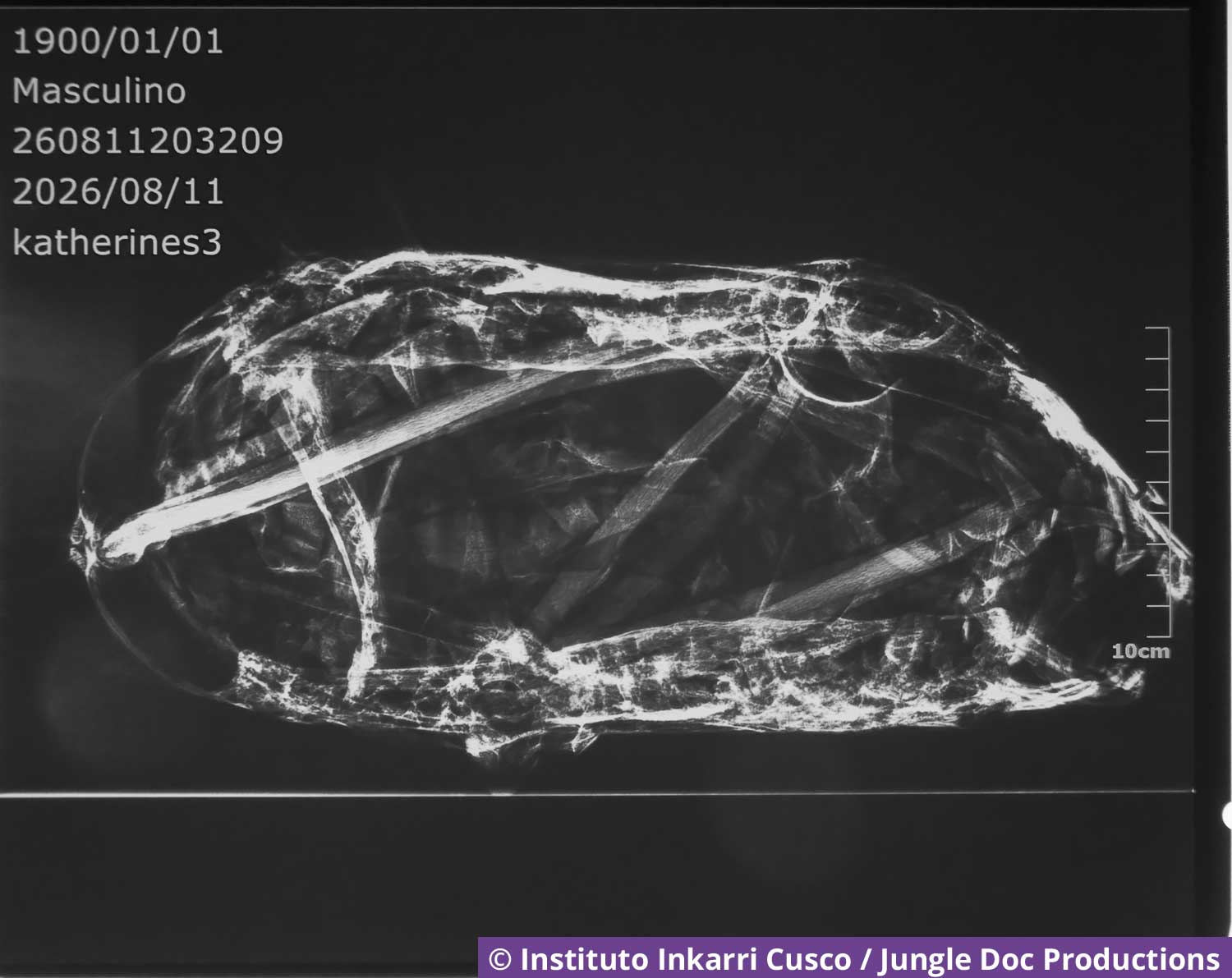
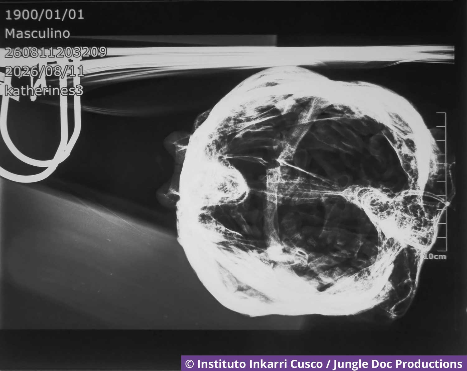
The Doctors' Doubts
Drs. Josep Valer and Edson Salazar Vivenco analyze Katarina's X-rays.
Observations
Christophe Serres, who developed the 3D Viewer, identified some interesting aspects based on Katarina's DICOM images.
Katarina's skull has a unique structure: it lacks the typical bony plates, but instead consists of “tubes” radiating from the eye sockets, with plates between these tubular structures.
- Tubular bone structures
- Plates between the tubular structures
- Four “spines.” Hollow bone structures that contain soft tissue
- Orbital structure, which is a key component of the skeletal structure
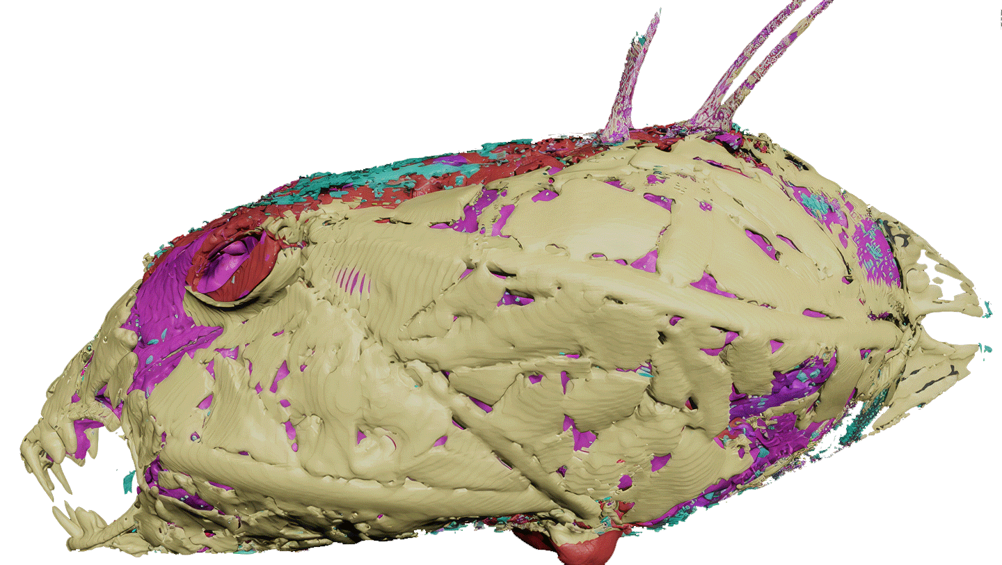
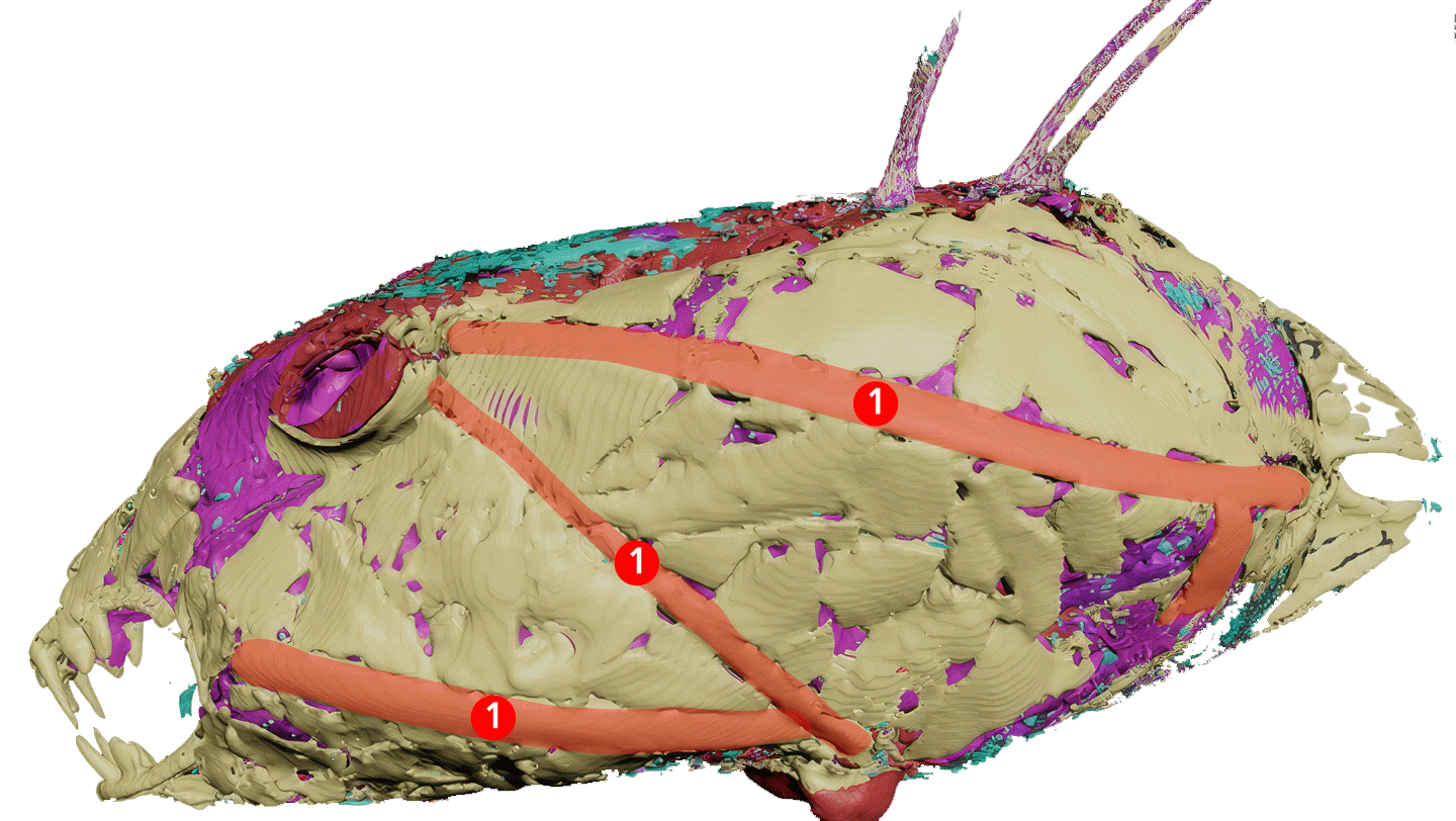
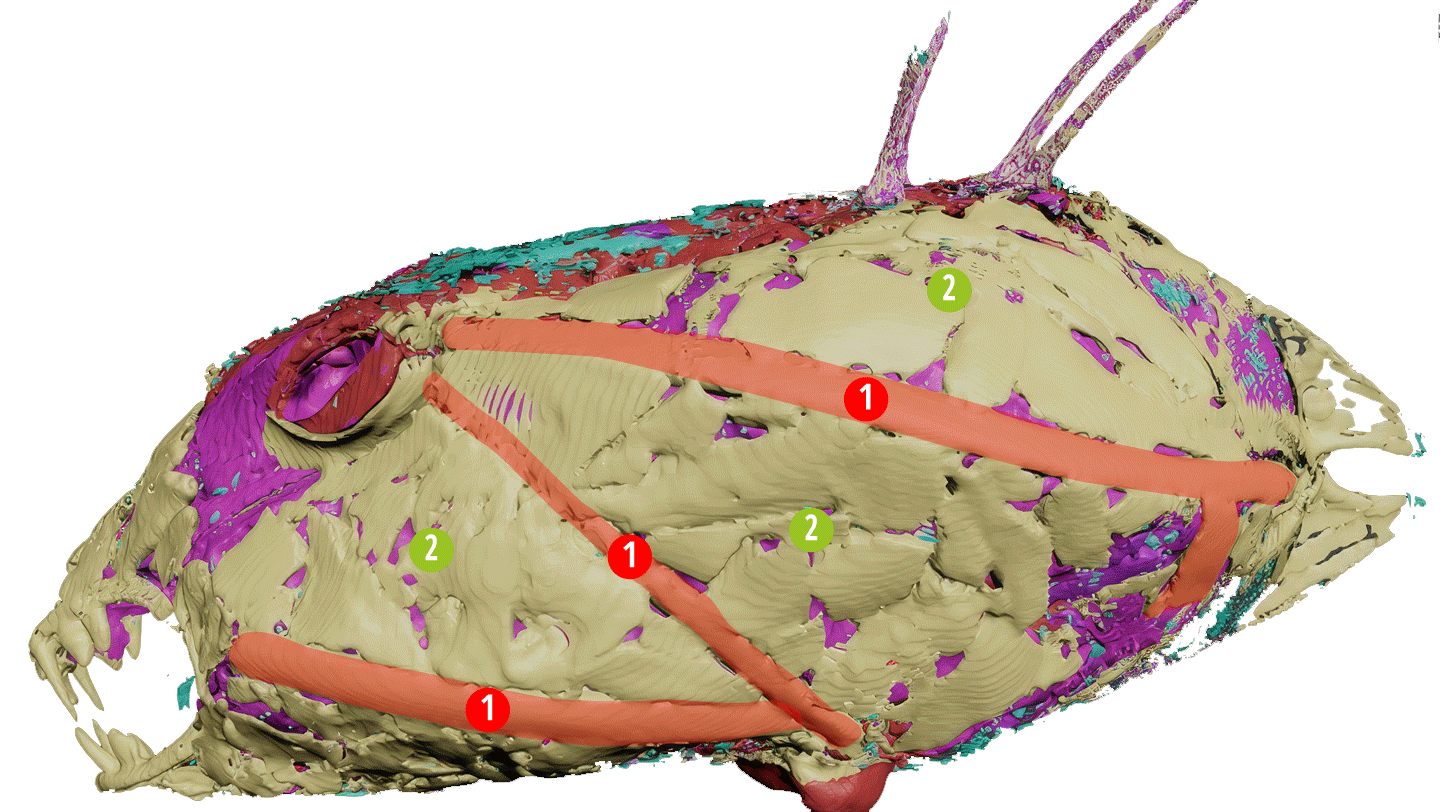
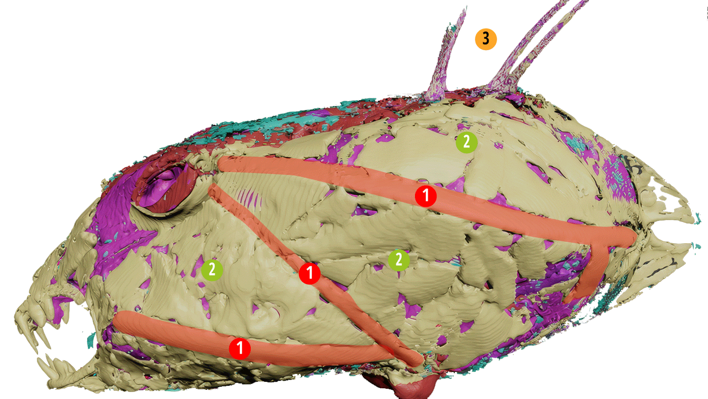
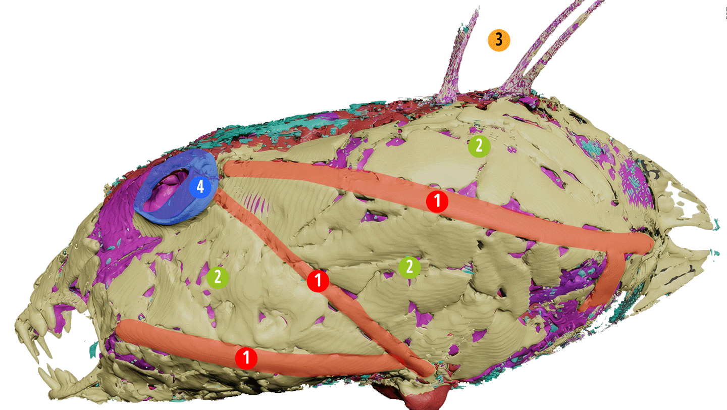
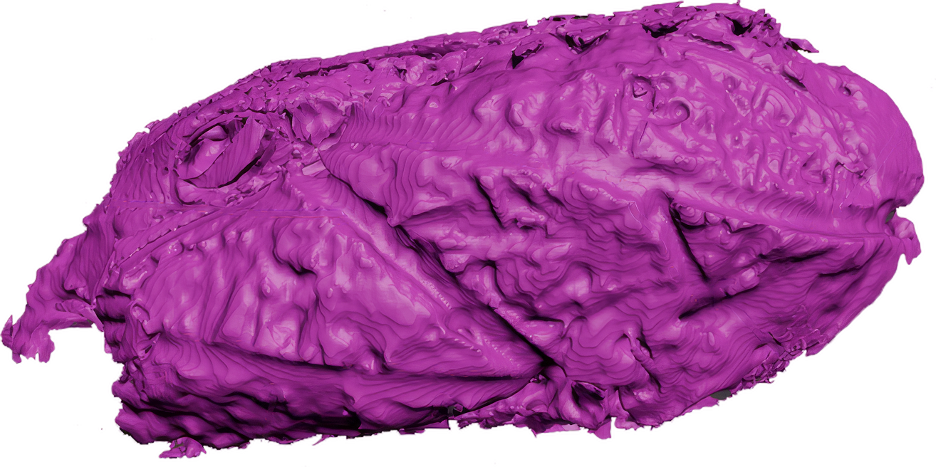
It is clear that the bone structure has left its mark on the muscles and soft tissues.
There appear to be two implants.
- The first is visible through the diatom layer.
- The second is deeply embedded in the epithelial layers, on the bony layer of the skull beneath the upper nostril.
Its shape and density suggest that it is an implant.
To find out more...

TRIDACTYLS The truth before us
52-page booklet
Buy

TRIDACTYLS Season 2
Documentary 6x60 min
Buy on VOD

TRIDACTYLS Season 1
Documentary 6x52 min
Buy on VOD


Lost Paititi & the Non-Human Remains of Nazca
English only
Buy